Is it a boy, is it a girl?
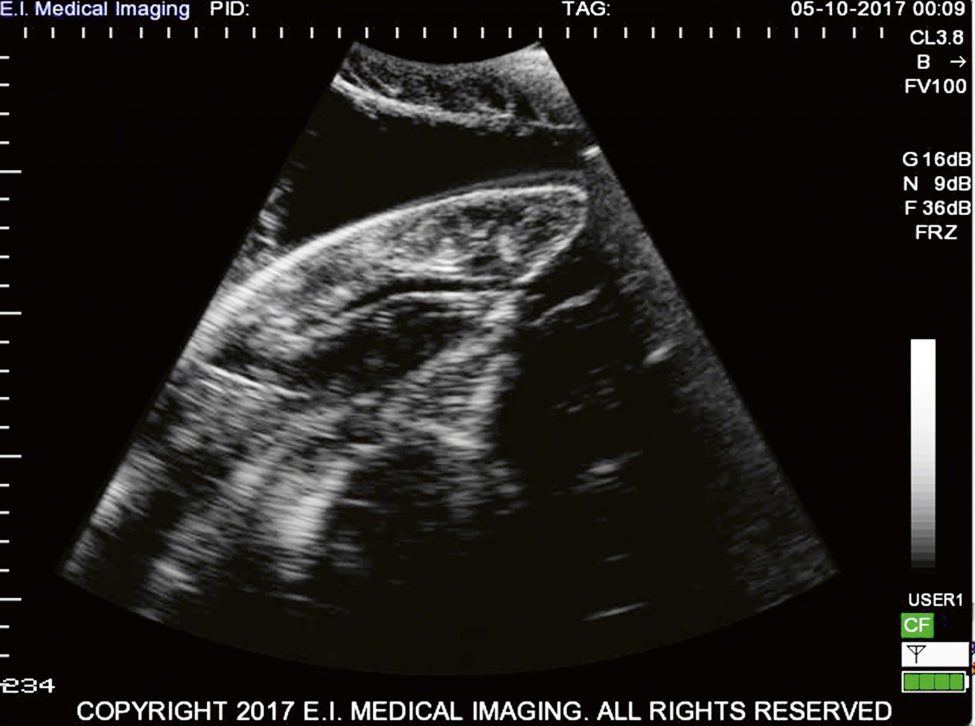
Sharks tend to keep their reproductive secrets close to their belly, which is why giving a tiger shark an ultrasound examination was such an amazing experience for the Shark Lab team.
Nursery grounds are important habitat for nearly all species. Locating such areas for wide-ranging animals can be difficult, especially in the marine environment where observing individuals is challenging. Within that environment, elasmobranchs form a group of approximately 1,000 members, which is relatively small compared to the more than 28,000 species of bony fish. Yet small though it may be, this group displays the most diverse means of reproduction: some members lay eggs, others give birth to live young – some even produce offspring by a combination of both. Skates deposit egg cases on the sea floor, sand tiger Carcharias taurus pups consume other embryos within the uterus, and lemon sharks Negaprion brevirostris are born with a placental connection to the mother similar to that of humans. Significantly, though, elasmobranchs produce relatively low numbers of offspring, which can be problematic for conservation efforts. Therefore it is important to understand their reproduction and the role it plays in their daily lives.
Advances in technology are providing non-invasive methods for assessing pregnancy; ultrasonography, for example, has been used for decades with humans. Incorporating this technology into shark research enables scientists to learn more about the reproductive attributes of individuals. This new information can be included in movement studies, population assessments and estimates of rebound potential.
Since the inception of the Bimini Biological Field Station (also known as Shark Lab) in 1990, our team has periodically encountered large female sharks with distended, rounded bellies. Most have been in the shallow-water long-line survey that we conduct each month, but some we have come across while scuba diving. Whenever these exciting moments occur, there’s one question that pops up in everyone’s mind and it quickly turns into a conversation on the boat or back at the lab: ‘Was that big female shark pregnant?’ ‘She must have been,’ comes the reply. ‘She was huge and her belly was prominent, possibly even moving. I’m sure she was shaped differently to a shark that has just fed!’ At least sharks are not offended by our suggestions!
Yet although our team was quietly confident that many of the big momma sharks it had encountered in the past were indeed pregnant, we had no way of confirming or proving it. This is, of course, a major problem for scientists, as we typically rely on several lines of evidence to convince colleagues and journals of our findings.
By a stroke of good fortune, in 2007 an expert in animal ultrasonography, who was working on a TV series called Into the Womb, visited the Shark Lab and used a field ultrasound to confirm the pregnancy of a huge adult lemon shark. This was a fascinating experience for the team and an important first insight into this developing technology. But it took another decade – in fact, until earlier this year – before we finally got our hands on a unit for daily use in our research. E.I. Medical, a leading manufacturer of ultrasound equipment, generously donated one of its machines for our use. This past year has been a steep learning curve with some exciting moments, one of which is described by the Shark Lab’s director, Dr Tristan Guttridge.
Bimini Biological Field Station (BBFS)
Samuel, better known as Doc, has been studying sharks for 50 years. He discovered how sharks see and even gave us insights into how they think. He founded the Bimini Biological Field Station in 1990, and has been training and inspiring young shark researchers ever since.


